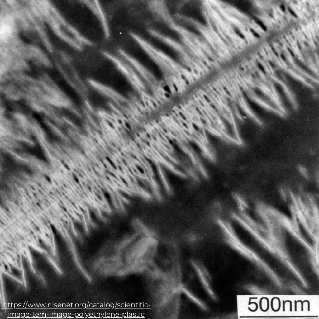
The electron microscope, due to the wave properties of emitted electrons from a thin filament of thermionic material (the material which has the flow of charged particles called thermions from a charged metal or a charged metal oxide surface, caused by thermal vibrational energy overcoming the electrostatic forces holding electrons to the surface) or a field emitter source, has a high resolving power that allows the observation of very small samples (1). Transmission electron microscopy (TEM) is commonly employed to characterize nanomaterials, providing chemical information and atomic-scale images. However, TEM is not suitable for visualizing nanoparticles (NPs) and polymers due to their amorphous structure. Heavy-metal stains are required to enhance contrast and visualize the microstructure of NPs and polymers (2).
TEM is not used for characterizing microplastics (MPs) but it is frequently utilized in studies examining the effects of MPs on model systems. For instance, Sun et al. (2018) investigated the toxic effects of polystyrene nano- and micro-plastics on the marine bacterium Halomonas Alkaliphila, examining growth inhibition, chemical composition, inorganic nitrogen conversion efficiencies, and generation of reactive oxygen species (ROS) using TEM (3). They observed increased extracellular polymeric substances, which may serve as possible bacterial protective mechanisms. In another study, TEM characterization was performed to evaluate the possible effects of MPs (PP, PE, PET, and PVC) on microalgae, analyzing growth inhibition and variations in cell structure (4). Other microscopic techniques like scanning electron microscopy (SEM), fluorescence microscopy, and atomic force microscopy (AFM) are preferred for characterizing MPs based on their size, surface characteristics, thickness, and other physicochemical properties.
The accuracy of visual identification of microplastics using TEM may vary depending on the user. To minimize errors, a well-established protocol can be employed to cross-validate results from multiple users. Additionally, further analysis using Energy-dispersive X-ray spectroscopy (EDS) can provide information on the elemental composition of microplastics, including additive components. EDS can also aid in distinguishing carbon-rich plastics from inorganic particles by analyzing the surface elemental composition. Nonetheless, the use of TEM, and SEM as well, is limited due to the expensive equipment required, as well as the time and effort involved in sample preparation and inspection (5).
Ref:
- Mariano S, Tacconi S, Fidaleo M, Rossi M and Dini L (2021) Micro and Nanoplastics Identification: Classic Methods and Innovative Detection Techniques. Front. Toxicol. 3:636640. doi: 10.3389/ftox.2021.636640
- D. Kalaronis, N.M. Ainali, E. Evgenidou, et al., 2022, “Microscopic techniques as means for the determination of microplastics and nanoplastics in the aquatic environment: A concise review”, Green Analytical Chemistry 3, 100036
- Sun, X., Chen, B., Li, Q., Liu, N., Xia, B., Zhu, L., et al. (2018). Toxicities of polystyrene nano- and MPs toward marine bacterium Halomonas alkaliphila. Sci. Total. Environ. 642, 1378–1385. doi: 10.1016/j.scitotenv.2018.06.141
- Song, C., Liu, Z., Wang, C., Li, S., and Kitamura, Y. (2020). Different interaction performance between MPs and microalgae: the bio-elimination potential of Chlorella sp. L38 and Phaeodactylum tricornutum MASCC-0025. Sci. Total. Environ. 723:138146. doi: 10.1016/j.scitotenv.2020.138146
- Woo, H.; Seo, K.; Choi, Y.; Kim, J.; Tanaka, M.; Lee, K.; Choi, J. Methods of Analyzing Microsized Plastics in the Environment. Appl. Sci. 2021,11,10640. https://doi.org/ 10.3390/app112210640
By: Moe Thazin Shwe, SOLEN Research Associate – IPC panel member
Editted by: Hendra WINASTU, SOLEN Principal Associate – IPC panel coordinator
Date: 10 May 2023
Article#: SOLEN-IPC-0017

 Tiếng Việt
 日本語
日本語
Pingback: Thermal Analysis for Microplastics Identification - Solen
Pingback: Atomic Force Microscopy and Nanoplastics - Solen
Pingback: Atomic Force Microscopy and Nanoplastics - SOLEN-IPC-019 - SOLEN
Pingback: 原子間力顕微鏡とナノプラスチック- SOLEN-IPC-019 - Solen